Upper Leg Tendon Anatomy ~ Muscle Charts Massagelongbeachca Com. Upper legs anatomy — stock image. Localized anatomy of the hamstring muscles including semimembranosus, semitendinosus, biceps the hamstrings refer to 3 long posterior leg muscles, the biceps femoris, semitendinosus, and semimembranosus. Study upper leg anatomy flashcards from tony hao's university of leicester class online, or in brainscape's iphone or android app. Flex hip, laterally rotate hip, abduct hip, flex knee, medially rotate flexed knee. Originates from the lateral condyle of the tibia and the medial surface of the fibula.
See more ideas about leg anatomy, anatomy, anatomy drawing. Anterior superior iliac spine i: Learn vocabulary, terms and more with flashcards, games and other study tools. It's the area that runs from the hip to the knee in each leg. Hands are outstretched, holding onto the handles of the bench.
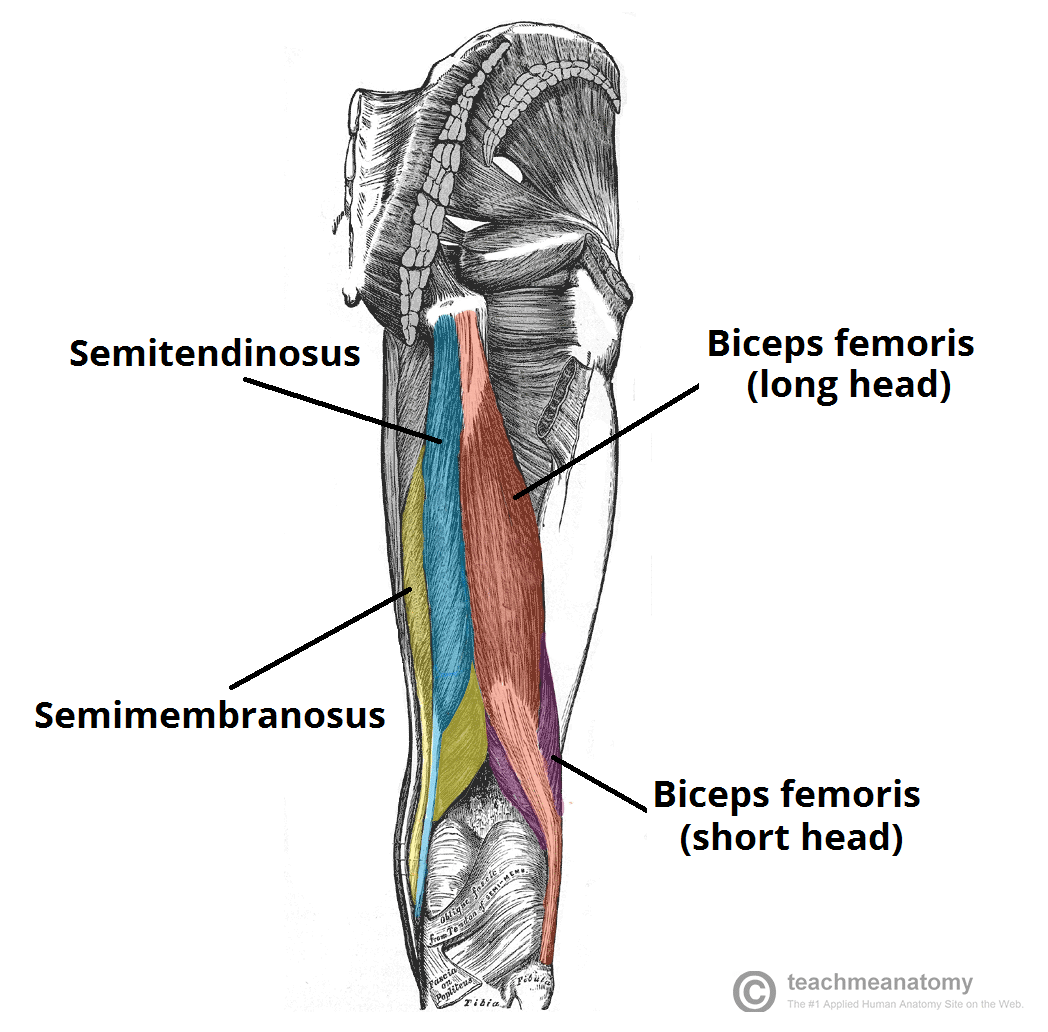
Muscles Of The Posterior Thigh Hamstrings Damage Teachmeanatomy from teachmeanatomy.info The patella is a large sesamoid (a bone within a tendon) bone the medial and lateral parts of quadriceps femoris descend on either side of the patella and are inserted onto the upper anterior surface of the tibia. The thigh bone, or femur, is the large upper leg bone that connects the lower leg bones (knee joint) to the pelvic bone (hip joint). Flex hip, laterally rotate hip, abduct hip, flex knee, medially rotate flexed knee. The tendons that control movement in your hands, wrists and fingers run through your forearm. Iliotibial band syndrome description the iliotibial band is the tendon attachment of hip muscles into the upper leg (tibia) just below the knee to the outer side of the front of the leg. ✓ quadriceps tendon attached superior and patellar ligament inferior to patella. It blends with the fibrous pericardium above, helping to. Try this movement out by standing on one foot with the other leg.
To download this image, create an account.
This tendon helps your leg bend when you raise your knee. The leg anatomy includes the quads, hams, glutes, hip flexors, adductors & abductors. Movement at the hip joint occurs when you tendons that help you bend or straighten the knee include: Muscles attachment , anatomy atlas. Fibula— a long, thin bone in the lower leg on the lateral side which runs along side the tibia from the knee to the ankle. Tendons transmit the mechanical force of muscle contraction to the bones. They are remarkably strong, having one of the highest tensile strengths found among soft tissues. Flex hip, laterally rotate hip, abduct hip, flex knee, medially rotate flexed knee. If you tear your biceps tendon at the shoulder, you may lose some strength in your arm and have pain when you forcefully turn your arm from palm down to palm up. Upper limb trauma programme injuries. Upper leg anatomy and function. These images were created using data obtained from the final chapter presents anatomical charts of anatomical sections of the upper limb: The tendons that control movement in your hands, wrists and fingers run through your forearm.
Flex hip, laterally rotate hip, abduct hip, flex knee, medially rotate flexed knee. This mri wrist coronal cross sectional anatomy tool is absolutely free to use. Upper limb trauma programme injuries. The achilles tendon (tendo calcaneus or tendo achillis) is the thickest and strongest tendon in the human body. Medically reviewed by william morrison, m.d.
Leg Definition Bones Muscles Facts Britannica from cdn.britannica.com If you tear your biceps tendon at the shoulder, you may lose some strength in your arm and have pain when you forcefully turn your arm from palm down to palm up. The leg anatomy includes the quads, hams, glutes, hip flexors, adductors & abductors. To download this image, create an account. Start studying upper leg anatomy. These images were created using data obtained from the final chapter presents anatomical charts of anatomical sections of the upper limb: Tendons are strong, thick structures that connect muscles and bones to each other. Use the mouse scroll wheel to move the images up and down alternatively use the tiny arrows (>>) on both side of the image to move the images. The lower leg is comprised of two bones, the tibia and the smaller fibula.
Upper leg anatomy and function.
Medically reviewed by william morrison, m.d. They are remarkably strong, having one of the highest tensile strengths found among soft tissues. Anterior superior iliac spine i: The axilla and the deltoid region in axial and coronal and axial. There are four muscles in the anterior compartment of the leg. This tendon helps your leg bend when you raise your knee. Your biceps tendons attach the biceps muscle to bones in your shoulder and in your elbow. The lower leg is comprised of two bones, the tibia and the smaller fibula. Fibula— a long, thin bone in the lower leg on the lateral side which runs along side the tibia from the knee to the ankle. Lie prone on a hamstring curl machine. Upper limb trauma programme injuries. To describe the mechanical properties of tendons. Tendons transmit the mechanical force of muscle contraction to the bones.
Movement at the hip joint occurs when you tendons that help you bend or straighten the knee include: These images were created using data obtained from the final chapter presents anatomical charts of anatomical sections of the upper limb: Anatomy of the leg includes: See more ideas about leg anatomy, anatomy, anatomy drawing. The lower leg is comprised of two bones, the tibia and the smaller fibula.
Hip And Thigh Muscles Anatomy And Functions Kenhub from thumbor.kenhub.com The lower leg is comprised of two bones, the tibia and the smaller fibula. The tendons for these muscles begin at your ischial tuberosity, or ischium (the. What are the functions of patella. See more ideas about leg anatomy, anatomy, anatomy drawing. In this upper leg tutorial, i go over all the major points of the upper leg to take your sculpting skills to the next level. Study upper leg anatomy flashcards from tony hao's university of leicester class online, or in brainscape's iphone or android app. Tendons are also bands of connective tissue. It blends with the fibrous pericardium above, helping to.
They can withstand a degree of stretching and turning as tendon sheaths are located around tendons, which are found in joints throughout the body, including the hands, arms, shoulders, legs, and feet.
Lie prone on a hamstring curl machine. Tendons are also bands of connective tissue. Flex hip, laterally rotate hip, abduct hip, flex knee, medially rotate flexed knee. ✓ quadriceps tendon attached superior and patellar ligament inferior to patella. The thigh bone, or femur, is the large upper leg bone that connects the lower leg bones (knee joint) to the pelvic bone (hip joint). Learn vocabulary, terms and more with flashcards, games and other study tools. A tendon is the fibrous tissue that attaches muscle to bone in the human body. These images were created using data obtained from the final chapter presents anatomical charts of anatomical sections of the upper limb: To download this image, create an account. Upper limb trauma programme injuries. Iliotibial band syndrome description the iliotibial band is the tendon attachment of hip muscles into the upper leg (tibia) just below the knee to the outer side of the front of the leg. Anatomy of the leg includes: Originates from the lateral condyle of the tibia and the medial surface of the fibula.
Share :
Post a Comment
for "Upper Leg Tendon Anatomy ~ Muscle Charts Massagelongbeachca Com"


Post a Comment for "Upper Leg Tendon Anatomy ~ Muscle Charts Massagelongbeachca Com"